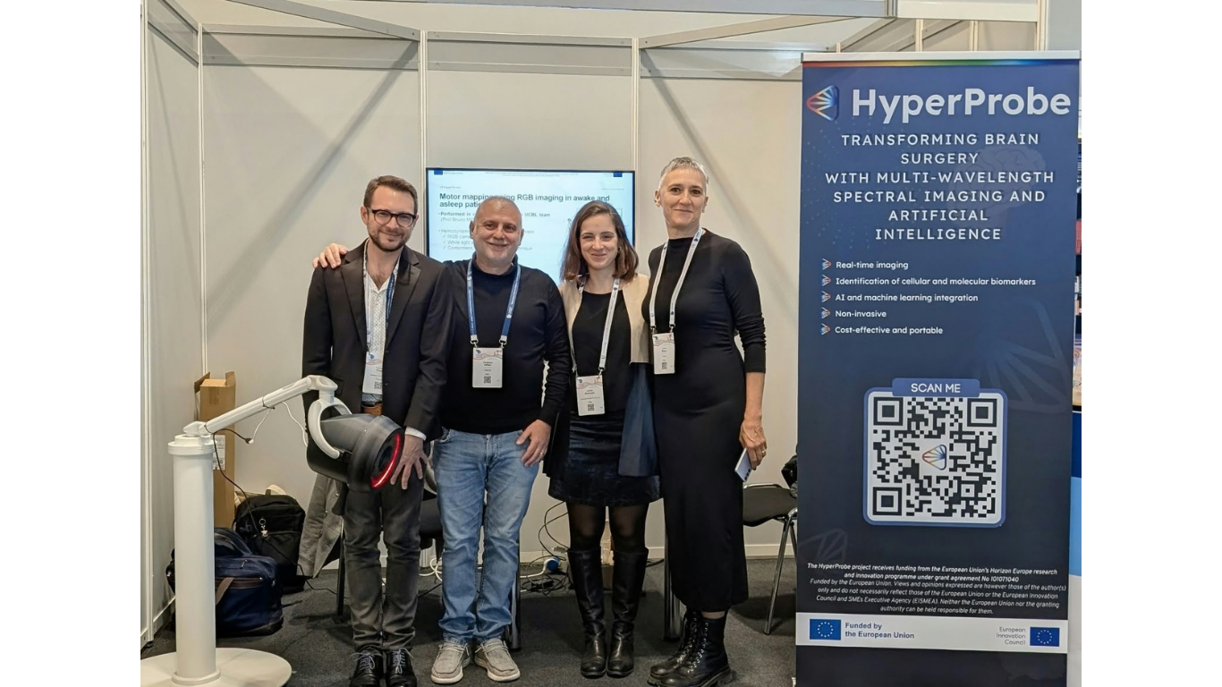
On March 6th, at the European Congress of Radiology, Pietro Ricci (The University of Florence) highlighted in a live session how the HyperProbe project could reshape neurosurgery, addressing long-standing clinical challenges with more precise, real-time insights.
The session focused on the central issue of brain tumor surgery and its difficulty of accurately identifying tumor boundaries and localization during the procedure. Surgeons often rely on limited intraoperative imaging, while tumor tissue can appear indistinguishable from healthy brain matter to the naked eye. Pietro Ricci presented HyperProbe’s solution: a hyperspectral imaging system capable of detecting oxygenated hemoglobin and mapping tissue oxygenation in real time. By analyzing molecular “imprints” rather than relying solely on visual cues, the technology enables differentiation between tumor types and surrounding tissue much faster than conventional methods. This is particularly relevant because prolonged oxygen deprivation alters vascular metabolism, information that can now be visualized intraoperatively. The result is improved margin delineation and a better chance of preserving critical brain function, especially given that brain tissue can shift during surgery and requires continuous feedback.
The overarching message of the HyperProbe session was clear: incremental advances in imaging are no longer enough. By combining hyperspectral imaging, photonics, and AI-supported interpretation, these technologies may offer a pathway toward more reliable diagnostics and safer, more precise interventions. Just as importantly, Pietro emphasized that successful adoption would depend on close collaboration between researchers, radiologists and clinicians to translate technical innovation into everyday clinical practice.
We are proud of our researchers’ work and dedication in advancing this technology.
Check out the presentation here!
Gallery Below